Story & photos by COMM 1316/News Photography Class
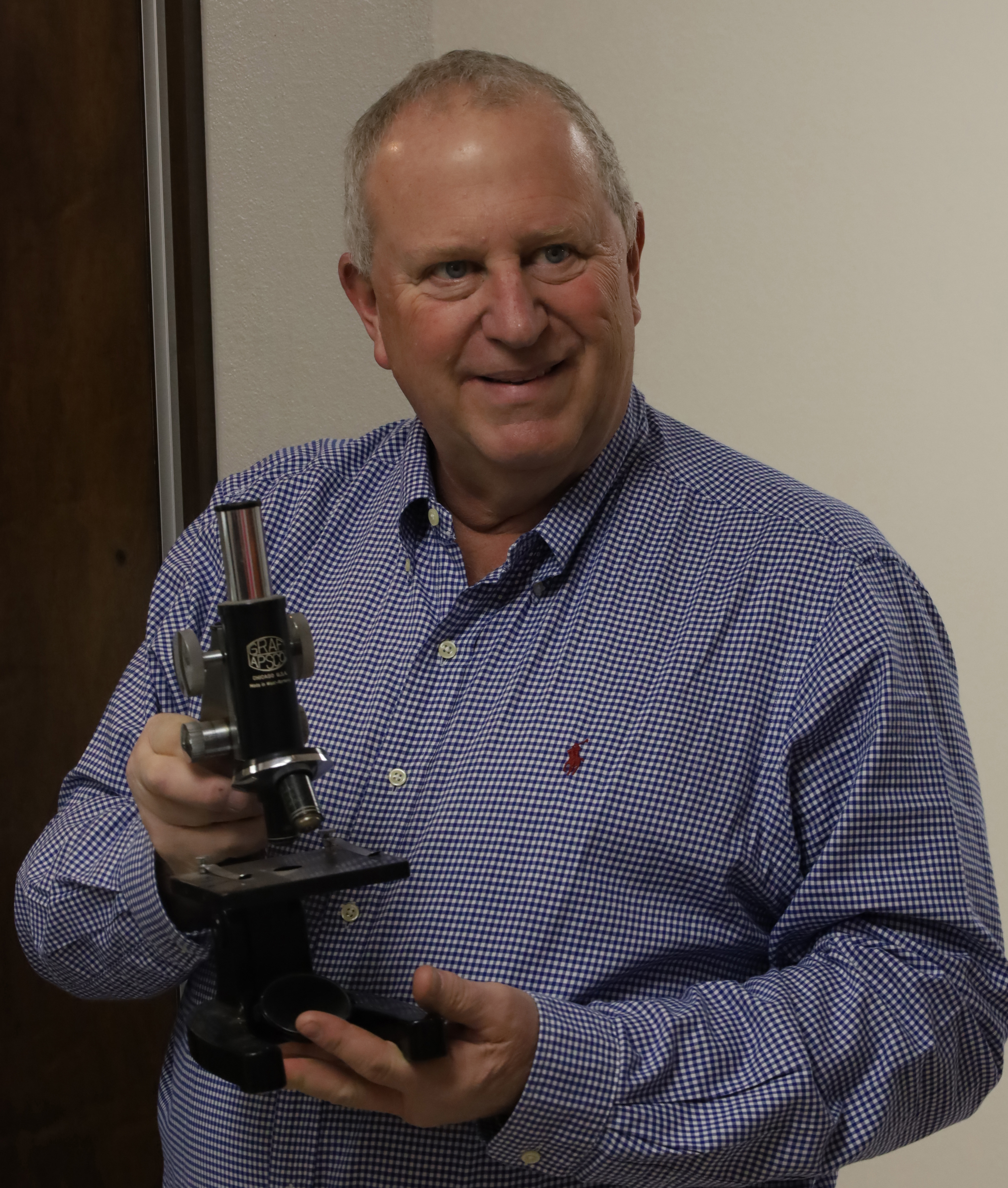
“I teach with microscopes,” Phil Ricker says. And Ricker has been teaching microbiology at South Plains College for 35 years now.
“Microbiology is the study of organisms that are so small, you have to have a microscope to see them,” he says.
Since Ricker uses microscopes so much, it’s no surprise really that he is the
person behind the display of antique models in the new Wilburn and Helen
Wheeler Science Center on the Levelland campus.
Thirty or so antique scopes, most are reproductions, are perched on shelves behind locked glass panels near the microbiology classrooms. Ricker showed them off recently for students in News Photography Comm 1316 so they could photograph them.

His favorite microscope, he says, is the oldest. It’s only about three inches tall and it’s made of shiny brass colored metal.

“It wasn’t made by a scientist,” he says. “It was made by a store merchant in the middle 1600s who wanted to see the quality of his fabrics.” He demonstrates how a little piece of fabric could be placed on the end, so when it’s held up to the light, a person could see the fabric’s thread count.


The next oldest models, he says, were not made for scientists either. “Starting
in the 1700s it became kind of a fad,” he says, “that people wanted to look at stuff under a microscope. So, they sold these for people to have at home. Because they were looking at a world that they didn’t know existed.”


Ricker describes another model that sits on the top shelf inside a wooden box. He says he likes this one because he was also able to get the newspaper ad for the scope when it was being sold sometime in the 1920s and 1930s.
“That cost $143 a 100 years ago,” he says. “That was a lot of money back then to be able to get a modern microscope.”

There was no light source on microscopes, Ricker explains, until probably the 1960s. Until then, in order to illuminate a slide, a person had to use the sun or a table light to catch light onto a mirror and reflect it up to the slide.


Ricker moved one of the old models into his microbiology classroom so photography students could compare it to a microscope a student would use in his class today.


The biggest difference between the old and new, he says, is the ability to adjust and focus with precision. In other words, it has to do with quality of the optics or the glass.


Photography students practiced pointing their cameras through the eyepiece of the modern scope to see if they could capture what was on the slide below. Most were able to snap pinkish red squiggles. Ricker says the squiggles are bacteria.


“Microscopes are used in all aspects of biology,” he says, “by the medical and dental field, pathologists use them to determine if cells are cancerous, and even the tech industry uses them to examine chips.”

Leave a Reply